Coronavirus today and the SCI community tomorrow

I’m not going to sugar coat the Coronavirus situation. Even as I begin writing this post, the numbers continue to grow geometrically for new coronavirus cases around the world. The day started out with around 460 cases in the USA but surpassed the 500 mark earlier than some experts expected. There are currently 538 coronavirus cases and 22 deaths in 32 states. Worldwide, the case count is 109,936. For those wanting to track the numbers worldwide, a good site to do that is https://www.worldometers.info/coronavirus/

For a dashboard glimpse from John Hopkins, https://www.arcgis.com/apps/opsdashboard/index.html#/bda7594740fd40299423467b48e9ecf6

It’s important to be prepared for the well being of yourselves and family members. The spread of this virus will not slow anytime soon, it will not miraculously disappear in warm weather. Depending on your location (I currently have readers in over 100 countries) the government response seems to be getting somewhat better. Most countries were not prepared for the speed or intensity of the COVID-19 outbreak so the measures have been slow and ineffective in stopping the rapid spread.

With the sheer numbers climbing at such a fast pace, the local health organizations, hospitals and clinics and will be pushed beyond their capabilities. The infrastructures were simply not in place. Test kits are very slowly making their way out (75,000 instead of the projected 1,000,000). There remains a shortage of equipment and supplies. There are simply not enough hospital beds for the cases that will be coming up and there are nursing shortages because of them also becoming ill. There will be no vaccine for another year or 18 months.

Be sure to stock up on several weeks of supplies. Much of your needs can be found and ordered online until the supply lines adjust.

World Health Organization LINK

Nurse Linda at the Christopher Reeve Foundation wrote a great piece about SCI, flu and coronavirus a few days ago. https://www.christopherreeve.org/blog/life-after-paralysis/the-flu

This was a good post today at CareCure by SCI Nurse (KLD)

“I don’t think we really know much about SCI/D and coronavirus yet, but what we do know is that people with SCI do have somewhat depressed immune systems to start with, and those with higher injuries who have less ability to cough and clear secretions are much more vulnerable to pneumonia, and also to ARDS, which can result from pneumonia.

Unfortunately, neither flu shots nor pneumonia immunizations provide any protection from the coronavirus. The pneumonia immunization does not prevent pneumonia from viruses (like the flu, or coronaviruses), but only from a number of bacterial causes of pneumonia.

Keep in mind that pneumonia is not caused by an specific infectious agent, but is actually a complication of a respiratory infection from either bacteria or viruses, which is much more likely to occur in those who are immuno-suppressed or have a decreased ability to cough up secretions.

At this time, the best prevention is to avoid being around people who could have been exposed to the coronavirus:

This may mean avoiding large crowded spaces, such as theaters, concert halls, cruise ships, airports, etc.
Be sure that your caregivers, if any, do not come to work with any signs or symptoms of respiratory infection (either the flu, bad cold, or cough or fever).
Have a back-up plan for care so you are not forced to have them come to work when sick.
Be sure family members and caregivers know how to properly sneeze and cough, and be meticulous about hand hygiene.
Washing hands properly with soap and water for at least 20 seconds is fine…reserve the use of hand sanitizer for places where there is no access to hand washing facilities and it should have 60% alcohol.
Maintain at least 5 feet distance from anyone you see in public who is coughing or sneezing.
Do not shake hands with anyone, and avoid hugging as well.
People with upper respiratory infections should wear masks. There is little evidence that wearing ordinary surgical masks (anything other than an N95 mask) will do anything for prevention of acquiring an infection, other than to keep you from touching your face, mouth, nose, or eyes with dirty hands.”
(KLD)

Posted in Uncategorized | Comments Off on Coronavirus today and the SCI community tomorrow

Stroke Drug Boosts Stem Cell Therapy For Spinal Cord Injury In Rats

Four months after treating them, Yasuhiro Shiga, MD, PhD, checked on his rats. Walking into the lab, he carried minimal expectations. Treating spinal cord injuries with stem cells had been tried by many people, many times before, with modest success at best. The endpoint he was specifically there to measure — pain levels — hadn’t seemed to budge in past efforts.

“Well, it doesn’t seem to be working. I don’t see any real change in pain behavior in any of the groups,” said Shiga, a visiting scholar at University of California San Diego School of Medicine, apologetically, as he walked into the office of his supervisor, Wendy Campana, PhD, professor in the Department of Anesthesiology and Program in Neuroscience.

But, to Campana’s surprise, he continued, almost as an after-thought.

“Although … some rats are actually really moving.”

The difference for those rats was this: Before delivering them into the spinal cord injury site, Shiga and Campana had conditioned stem cells with a modified form of tissue-type plasminogen activator (tPA), a drug commonly used to treat non-hemorrhagic stroke.

Read the Full Article in ScienMag

UC San Diego News Center Article: By Heather Buschman, PhD

Their findings are published December 17, 2019 in Scientific Reports.

Nature Publication Authors: Yasuhiro Shiga, Akina Shiga, Seiji Ohtori, Chiba University; Pinar Mesci, HyoJun Kwon, Coralie Brifault, John H. Kim, Jacob J. Jeziorski, Chanond Nasamran, Alysson R. Muotri, and Steven L. Gonias, UC San Diego.

Posted in Regenerative Medicine, spinal cord injury research, Stem Cell Research | Tagged , , , , , , , , , , | Comments Off on Stroke Drug Boosts Stem Cell Therapy For Spinal Cord Injury In Rats

An integrated in silico pipeline identifies a novel TF combination that promotes enhanced CST growth following injury

Authors
*I. VENKATESH1, Z. WANG2, V. MEHRA1, E. EASTWOOD1, M. SIMPSON1, A. CHAKRABORTY1, D. GROSS1, Z.BEINE1, M. CABAHUG1, G. OLSON1, M. G. BLACKMORE3;2Dept. of Biomed. Sci., 3Biomed. Sci., 1Marquette Univ., Milwaukee, WI

Ishwariya Venkatesh PhD Marquette University

Lab Abstract
Embryonic and peripheral neurons respond to axonal injury with activation of transcriptional networks conducive to re-growth. In contrast, injured mature CNS neurons fail to re-induce appropriate transcriptional networks, resulting in failed regeneration and permanent damage. We have previously shown that forced re-expression of single transcription factors (TFs) such as KLF6 promotes axon outgrowth following injury and is a promising strategy for therapeutic neural repair. However, with single TF treatments the overall number and regenerative speed of axons remains sub-optimal, and likely insufficient for full functional recovery. Because TFs rarely function in isolation, we hypothesized that supplying combinations of TFs that synergize with KLF6 may boost growth phenotypes. To this end, we developed a bioinformatics pipeline to detect TFs that may synergize with KLF6 to drive the expression of pro-growth genes. To validate our bioinformatic predictions, we next systematically co-expressed candidate TFs in combination with KLF6 in assays of neurite outgrowth in post-natal CNS neurons. Remarkably, nearly 20% of the TF synergized with KLF6 to promote neurite outgrowth, validating the bioinformatics approach. To prioritize TFs for in vivo testing, we performed TF – target gene network analyses that identified 4 core TFs – EOMES, RARB, NKX32 and NR5A2 that are predicted to be critical to the regulation of pro-growth gene networks. Finally, we tested in vivo the ability of the 4 core TFs to promote CST axon growth individually or in combination with KLF6 following pyramidotomy injuries. Individually, RARB overexpression lead to increased CST sprouting following pyramidotomy. Importantly, we observed that although NR5A2 had no effect on growth by itself, forced co-expression with KLF6 promoted a robust increase in midline crossing by transduced CST axons, significantly above the level of KLF6 alone. Ongoing experiments are aimed at clarifying the molecular mechanisms underlying KLF6/NR5A2 synergy in driving axon growth. Overall, we describe a novel bioinformatics-based approach that has identified a completely novel TF combination that drives enhanced sprouting in the injured CST.

Grant Support
R21NS106309
R01NS107807
R01NS083983
Craig Nielsen Post doc fellowship (Venkatesh)

Posted in Chronic Spinal Cord Injury Research, Gene Therapy, Neuroscience Abstracts, spinal cord injury research | Tagged , , | 1 Comment

Spike timing-dependent plasticity in the adult rat with chronic cervical spinal cord contusion

Authors: *N. DE LA OLIVA1, A. E. HAGGERTY1, M. A. PEREZ1,2, M. OUDEGA1,2,3,4;
1 Miami Project To Cure Paralysis, Univ. of Miami, Miami, FL;
2 Bruce W. Carter Dept. of Veterans Affairs Med. Ctr., Miami, FL;
3 Affiliated Cancer Hosp. & Inst. of Guangzhou Med.Univ., Guangzhou, China;
4 Neurolog. Surgery, Miller Sch. of Medicine, Univ. of Miami, Miami,FL

Lab Abstract: Spinal cord injury (SCI) damages descending and ascending axons resulting in motor and sensory function impairments. Histological and electrophysiological data revealed that in most SCI patients residual axonal connections between the brain and the spinal cord below the injury exist, which opens avenues for neuromodulatory therapies for recovering function. electrical stimulation of residual axonal connections is a promising strategy to recover lost function. Repetitive electrical stimulation results in persistent increase or decrease of synaptic efficacy (i.e., long-term potentiation or depression, respectively). Previous studies demonstrated that the arrival of repeated pairs of precisely timed presynaptic and postsynaptic action potentials to a given synapse changes synaptic strength. This process is known as spike timing-dependent plasticity (STDP). The direction of the effects of STDP stimulation protocols depends on the spike order and time between the central and peripheral stimuli, as well as on the frequency and duration of the stimulation. Importantly, it was shown that STDP protocols can enhance motor function after paired corticospinal tract (CST) and peripheral nerve stimuli in people with and without SCI, although with transient effects. In this study, we aimed to elucidate the cellular and molecular mechanisms underlying STDP aftereffects in a cervical SCI rat model. First, we traced the CST and the reticulospinal tract (RST), which are both involved in forelimb reach and grasp behavior in rats, along with the motoneurons of targeted forelimb muscles, to evaluate the spinal connections before and after 12 weeks C5 chronic injury. Based on these results, an STDP stimulation protocol was applied to maximize the synaptic strength in those connections. Electrophysiological and histological techniques were used to evaluate changes after the stimulation. We hypothesize that higher frequency and longer stimulation will result in longerlasting functional and cellular aftereffects. The ultimate goal is to use the data from our animal studies to improve the efficacy of STDP protocol on improving function in SCI patients.

Program #/Poster #: 051.18/H4
Topic: C.11. Spinal Cord Injury and Plasticity
Support: VA Grant I01BX007080
Society for Neuroscience

Posted in Chronic Spinal Cord Injury Research, Neuromodulation, Neuroscience Abstracts, Rehabilitation, spinal cord injury research | 1 Comment

Effect of PTEN antagonist peptide on the functional motor recovery in rat

Authors: *S. LV1, W. WU2;
1 Guangdong-hongkong-Macau Inst. of CNS Regeneration, Guangzhou, China;
2 The Univ.Of Hong Kong, Hong Kong SAR, Hong Kong

Lab Abstract: Ventral root injury results in great loss of motor functions because of the inefficient axon regeneration and severe atrophy of target organ. PTEN act as a negative regulatory factor at PI3K/AKT pathway also inhibit the regeneration of axons. It has been shown that PTEN antagonist peptides(PAPs) can significantly stimulated growth of descending serotonergic fibers and sprouting of corticospinal fibers in the rostral spinal cord after spinal cord injury.

Here, we are reporting that after a spinal ventral root crush completely in adult rats, PAPs peptides treatment remarkably improved motor functional recovery. PAPs-treated animals showed less motoneuron death, increased the number of regenerated axons, rebuilt healthy neuromuscular junction and enhanced potentiated electrical responses of motor units. Our study showed that PAPs was a promising pharmacological method for promoting motor functional recovery after peripheral nerve injury.

Society for Neuroscience 2019
051. Axon Injury and Recovery
Location: Hall A
Time: Saturday, October 19, 2019, 1:00 PM – 5:00 PM
Program #/Poster #: 051.03/G33
Topic: C.11. Spinal Cord Injury and Plasticity

Posted in Chronic Spinal Cord Injury Research, Neuroscience Abstracts, Regenerative Medicine, spinal cord injury research | 1 Comment

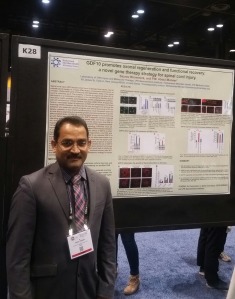
GDF10 promotes axonal regeneration and functional recovery: A novel gene therapy strategy for spinal cord injury

PM Abdul Muneer PhD at Society for Neuroscience 2019 Poster Presentation


Authors:*P. M. ABDUL-MUNEER, S. BHOWMICK, V. D’MELLO;
Hackensack Meridian Hlth. JFK Med. Ctr., Edison, NJ
Neuroscience 2019 LINK

Lab Abstract: Spinal cord injury (SCI) occurs when there is damage from trauma, loss of normal blood supply, or a mass effect due to compression from tumor or infection. Unlike other parts of the body, the regenerative ability of the spinal cord is relatively poor. The inability of axons to regenerate after SCI is attributable to a combination of effects of the non-permissive extrinsic factors including myelin proteins and chondroitin sulfate proteoglycans (CSPGs), and cell-autonomous intrinsic factors including cAMP, RhoA, Krüppel-like factors, mammalian target of rapamycin (mTOR) and phosphatase and tensin homolog (PTEN). However, the factor(s) that may be triggered to promote the initiation of a molecular growth program and axonal sprouting in SCI are largely unknown. In this project, we developed a novel therapeutic approach to treat SCI by exploiting the neuronal growth-promoting potential of growth differentiation factor 10 (GDF10), a potential gene belongs to the transforming growth factor beta (TGF-β) superfamily. GDF10 regulates several molecular signaling systems to induce a neuronal growth state. Our focus on GDF10 as a therapeutic target after SCI is based on the observation that GDF10 regulates major axonal regenerative cues including PTEN, phosphoinositide 3-kinase (PI3K) and suppressor of cytokine signaling 3 (SOCS3). Thus, we hypothesize that up-regulation of GDF10 mitigates PTEN-mediated inhibition of axonal regeneration. We examined the specific effects of GDF10 on other major regulatory signaling cascades of axonal regeneration, the PI3K, and SOCS3 pathways in vitro and in vivo. In order to up-regulate GDF10 in experimental animals, we delivered GDF10 gene via adeno-associated virus into the sensory-motor cortical area of the brain and into the spinal cord rostral to the SCI lesion, and evaluate the subsequent progress of axonal regeneration and functional recovery after SCI. To validate the role of GDF10 in axonal regeneration, we used the CRISPR/Cas9 gene deletion technology to remove GDF10 gene. Findings from this project would help to clarify the specific role of GDF10 in axonal regeneration and functional recovery after SCI and establish a basis for pursuing GDF10 as a therapeutic strategy for spinal cord injured patients.

Grant Support:
NJ Commission on Spinal Cord Research Grant No. CSCR18ERG007
JFK Neuroscience Institute support package

Posted in Chronic Spinal Cord Injury Research, Gene Therapy, Neuroscience Abstracts, spinal cord injury research, Stem Cell Research | Tagged , , | 1 Comment

GTX Medical and NeuroRecovery Technologies to merge

GTX Medical and NeuroRecovery Technologies today announced their merger into a global company for the development of neuromodulation therapies for spinal cord injuries.

The two merging companies plan to develop the targeted epidural spine stimulation system, an implantable spinal cord stimulation platform designed to restore locomotion in patients with spinal cord injury with real-time motion feedback.

There is also a second, non-invasive product in the works, as a transcutaneous spinal cord stimulation system is in development for the restoration of upper limb movement and hand function.

SEE THE FULL ANNOUNCEMENT AT THIS LINK:

Posted in Chronic Spinal Cord Injury Research, Neuromodulation, Rehabilitation, spinal cord injury research | Tagged , | 3 Comments

Candidate Therapy From Quebec for Chronic SCI Being Developed in Parallel by Academics and Companies in Switzerland and the Netherlands

QUEBEC CITY, CANADA, October 10th 2019 – Today, Laboratoires Guertin announces that a campaign for funding has been launched to support a phase IIb-enabling pilot study with a tritherapy candidate (buspirone/L-DOPA/carbidopa)called Spinalon. This experimental oral pill has been shown to trigger short episodes of rhythmic leg activity in volunteers suffering chronically a severe spinal cord injury. In 2005, Spinalon was discovered as a pharmacological approach capable of eliciting, within minutes post-administration, spinal network activation and basic weight-bearing stepping on a treadmill for 30-45 minutes in completely paraplegic animals. In 2009, Nordic Life Science Pipeline signed an in-licensing agreement with Université Laval and Dr Pierre A. Guertin, the inventor, for the rights of testing SpinalonTM in a first-in-patient phase I/IIa study (NCT01484184). With support from the US Department of Defense (grant number W81XWH-11-1-08178) that first study was successfully completed in 2016 at the McGill University Health Center in Canada (Radhakrishna et al. Curr Pharm Des 23, 2017). Given that preliminary evidence of efficacy was found in completely paralyzed people, Laboratoires Guertin has decided to begin seeking funds for a pilot phase IIb enabling study in Belgium designed to demonstrate clinically-relevant efficacy on a treadmill in combination or not with medical devices such as muscle vibrators and exoskeletons. In parallel, Prof Dr Bloch, neurosurgeon in Switzerland (Centre Hospitalier Universitaire Vaudois), associate professor(Université de Lausanne), and co-founder of GTX Medical (Switzerland and The Netherlands) has announced, without authorization from Laboratoires Guertin and its founder, a clinical trial with buspirone/L-DOPA/carbidopa (SpinalonTM) in spinal cord-injured patients (NCT04052776).

Pierre Guertin PhD

About SPINALON
Discovered at Université Laval (Canada) in 2005 by Dr Pierre A. Guertin (full professor at the faculty of medicine), this proprietary tritherapy candidate (WO2006026850) constitutes a novel class of treatment acting as a potent activator of the spinal locomotor network also known as the central pattern generator (CPG) for locomotion (Guertin, Ung, Rouleau Biotechnol J 5, 2010). It is composed of already known and regulatory approved molecules for patients with Parkinson’s disease or anxiety. This drug treatment candidate was shown in paraplegic animals when used regularly (e.g., 3-5 times weekly) in combination with pharmacologically-enabled treadmill training to prevent or reduce the secondary complications and co-morbid problems associated with chronic paralysis (cardiovascular, metabolic, musculoskeletal, hormonal, and psychological problems) Guertin, Ung, Rouleau, Steuer Neurorehabil Neural Repair 25, 2011; Ung, Rouleau, Guertin, Neurorehabil Neural Repair 26, 2012).

About Laboratoires Guertin
Founded by Dr Pierre A. Guertin, it is an emerging specialty company based in Quebec with subsidiary companies based in Canada and Europe that focuses on the development of clinically-relevant pharmaceutical and biocosmeceutical products as well as of specialized literature and services in the field of chronic secondary consequences and co-morbidities associated with paralysis, bowel and bladder problems, reproductive dysfunction, and aging diseases such as cancer and metabolic disorders.
About Biosynergis Canada It is a subsidiary company of Laboratoires Guertin with a branch in Luxembourg (Biosynergis International) which mission is to focus on North America-based and Europe-based research activities, respectively, as well as on commercialization and sales worldwide of products developed by Laboratoires Guertin.

Posted in Chronic Spinal Cord Injury Research, Neuromodulation, Rehabilitation, spinal cord injury research | Tagged | 6 Comments

Moving beyond the glial scar for spinal cord repair

Authors: Elizabeth J. Bradbury and Emily R. Burnside
Abstract: Traumatic spinal cord injury results in severe and irreversible loss of function. The injury triggers a complex cascade of inflammatory and pathological processes, culminating in formation of a scar. While traditionally referred to as a glial scar, the spinal injury scar in fact comprises multiple cellular and extracellular components. This multidimensional nature should be considered when aiming to understand the role of scarring in limiting tissue repair and recovery. In this Review we discuss recent advances in understanding the composition and phenotypic characteristics of the spinal injury scar, the oversimplification of defining the scar in binary terms as good or bad, and the development of therapeutic approaches to target scar components to enable improved functional outcome after spinal cord injury.

Emily Burnside

Dr. Elizabeth Bradbury

Read the FULL ARTICLE at Nature Communications Volume 10, Article number: 3879 (2019)

Posted in Chronic Spinal Cord Injury Research, Regenerative Medicine, spinal cord injury research | Tagged , | 1 Comment

The Struggle to Make CNS Axons Regenerate: Why Has It Been so Difficult?

Author: James W. Fawcett
Abstract:
Axon regeneration in the CNS is inhibited by many extrinsic and intrinsic factors. Because these act in parallel, no single intervention has been sufficient to enable full regeneration of damaged axons in the adult mammalian CNS. In the external environment, NogoA and CSPGs are strongly inhibitory to the regeneration of adult axons. CNS neurons lose intrinsic regenerative ability as they mature: embryonic but not mature neurons can grow axons for long distances when transplanted into the adult CNS, and regeneration fails with maturity in in vitro axotomy models. The causes of this loss of regeneration include partitioning of neurons into axonal and dendritic fields with many growth-related molecules directed specifically to dendrites and excluded from axons, changes in axonal signalling due to changes in expression and localization of receptors and their ligands, changes in local translation of proteins in axons, and changes in cytoskeletal dynamics after injury. Also with neuronal maturation come epigenetic changes in neurons, with many of the transcription factor binding sites that drive axon growth-related genes becoming inaccessible. The overall aim for successful regeneration is to ensure that the right molecules are expressed after axotomy and to arrange for them to be transported to the right place in the neuron, including the damaged axon tip.

Read the Full Article at Neurochem Research

Posted in Chronic Spinal Cord Injury Research, Gene Therapy, Ligand, spinal cord injury research, Stem Cell Research | Tagged | Comments Off on The Struggle to Make CNS Axons Regenerate: Why Has It Been so Difficult?